

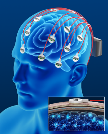
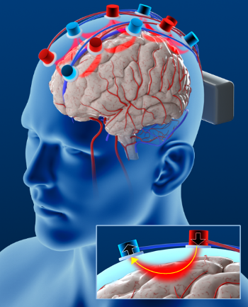
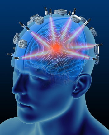
The brain activities reflect our cognitive, physiological, and psychological conditions. In CenBRAIN, we study various techniques focusing on multimodal neuroimaging systems for real-time brain activity monitoring. We explore the applications of multimodal functional near-infrared spectroscopy (fNIRS) and electroencephalogram (EEG) for diagnosis and prediction of neurodegenerative diseases, Figure 1a, b. In addition, photoacoustic imaging (PAI) is another emerging neuroimaging technique in our group. Figure 1c.
(a)
(b)
(c)
Figure 8. Neuroimaging techniques: (a) EEG, (b) fNIRS and (c) PAI.
One combination is integrating fNIRS and EEG techniques for brain activities monitoring [1]. EEG records the electrical activities of neural activities while fNIRS reflects the hemodynamic states of cerebrovascular. Hemodynamic states mean the variations of oxygenated and deoxygenated hemoglobin in blood vessels. At CenBRAIN, protocols are designed to study the changes of electrical and hemodynamic signals during various physical conditions, such as performing memory task, mental arithmetic, under mental stress or during imaginary condition. Collected signals indicating the changes of neural activity and neurovascular coupling caused by the stimulations. Moreover, these recordings are promising to be used for clinical applications, such as diagnosis, monitoring, outcome, onset events detection, and earlier prediction.
Photoacoustic imaging (PAI) is an emerging optical imaging technique. In CenBRAIN, we are investigating the multimodal stroke prediction system [2]. Intended for vascular imaging based on endogenous contrast from blood hemoglobin, PAI holds great potential to describe the cerebrovascular condition for stroke prediction. PAI modality uses optical absorption contrast and ultrasound resolution. Typically, PAI effect is produced by a short laser pulse. The pulse energy is partially absorbed by the tissue and converted into heat. Following a local transient temperature rise, the pressure rises through thermoelastic expansion. The pressure propagates, termed PA waves, and is detected by ultrasonic transducers. With high contrast, high spatial and time resolution, PAI is available to acquire 2D or 3D images to monitor the hemodynamics and metabolism in the brain. At present, we are exploring the optical and ultrasonic signals transmission to achieve multimodality for brain imaging.
[1] Y.-H. Chen and M. Sawan, "Trends and Challenges of Wearable Multimodal Technologies for Stroke Risk Prediction," Sensors, vol. 21, no. 2, 2021, Art no. 460, doi: 10.3390/s21020460.
[2] X. Yang, Y.-H. Chen, F. Xia, and M. Sawan, "Photoacoustic imaging for monitoring of stroke diseases: A review," Photoacoustics, vol. 23, p. 100287, 2021, doi: 10.1016/j.pacs.2021.100287.