The remarkable capacity of the human brain to perform complex functions relies on the integration and coordination of neural circuits across various cortical regions. At the core of this intricate network are inter-regional cortical tracts, which serve as fundamental architectural elements for connecting and orchestrating neural activity between distinct brain regions. These tracts are composed of axons that extend across regions, forming bundles known as cerebral tracts within the white matter. Extensive research on brain anatomy and connectivity, known as connectome studies, has revealed that a significant portion of axons in cerebral tracts establish reciprocal connections between two cortical regions. Specifically, association tracts are responsible for reciprocal interconnections within a single cerebral hemisphere. Understanding the mechanistic significance of these inter-regional projections in the development of neural circuits is a crucial step towards unraveling the mysteries of brain function.

Fig.1. Schematic representation of the wire-embedded 3D neural spheroid culture device, consisting of glass tube, agarose, PDMS and Pt wire electrode. The agarose provided a low adsorption surface to form neural spheroid and pre-inserted Pt wire electrode obtain interior signals recording without damage. The device allows for electrophysiology study from both interior and exterior of the neural spheroid and building neural tract connection model by employing simultaneous fading sinusoidal signals to stimulate two close neural spheroids. This platform has the potential to be expanded for use in various applications, including the internal supply of nutrients, real-time sensing, and organoids-based multi-brain region connection models.
A recent contribution from the CenBRAIN Neurotech, published in the Cyborg and Bionic Systems. In this paper, we introduce a novel 3D cell culturing and non-invasively characterization technique of neural spheroids.The PhD student Hongyong Zhang from our center is the first author of this work, Dr. Mohamad Sawan and Dr. Sumin Bian are the corresponding author. This work was supported by Zhejiang Key Laboratory of 3D Micro/Nano Fabrication and Characterization.
Reference:
ZHANG H., HUANG N., BIAN S., SAWAN M., “Platinum Wire-embedded Culturing Device for Interior Recording from Lollipop-shaped Neural Spheroids”, Online in the Cyborg and Bionic Systems Journal, 2025.
More information can be found at the following link:
https://spj.science.org/doi/10.34133/cbsystems.0220#tab-citations
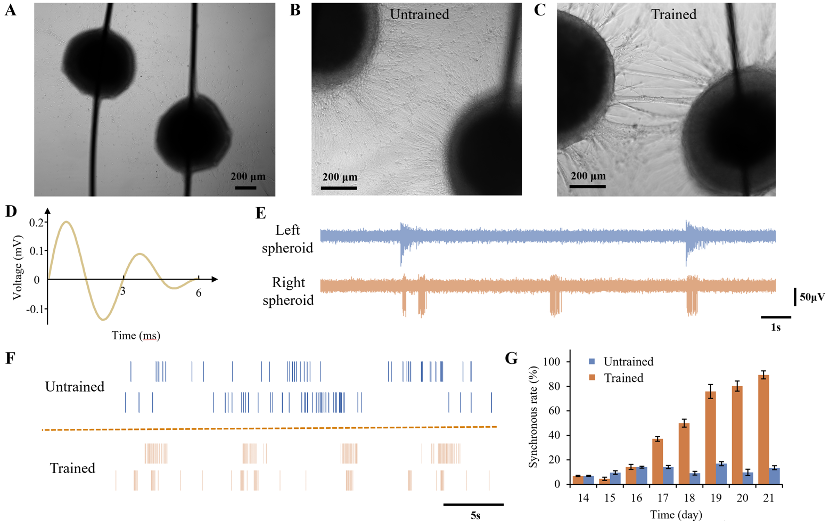
Fig. 2. Interconnection and synchronization between neural spheroids: A. Two lollipop-shaped neural spheroids positioned in close proximity, each extending axons towards the other; B. Axons connection between two neural spheroids without training, showing disorganized and sparse axonal; C. Axons connection between two neural spheroids with training, demonstrating focused and robust neural tract formation; D. Stimulation protocol: a fading sinusoidal signal with an amplitude of 0.2 mV and a period of 3s applied to the neural spheroids; E. Original neural signals recorded from the two trained neural spheroids, illustrating the quality of interior recordings; F. Raster plot comparing spontaneous firing patterns of the neural spheroids with and without training, revealing synchronized activity in the trained group; G. Synchronous firing rates of the neural spheroids measured on day 14 (start of the training) and subsequent days, showing enhanced synchronization with training.
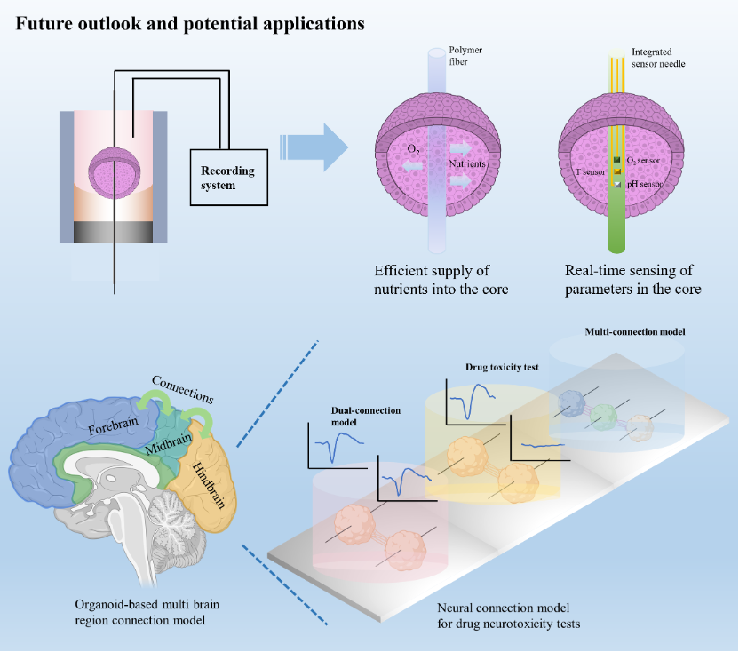
Fig. 3. Potential applications of the wire-embedded 3D neural spheroids culture device: First, replacing Pt wire with a porous polymer fiber could enable oxygen and nutrients to permeate the neural spheroids or brain organoids, potentially alleviating central hypoxia. Alternatively, using functional needles instead of Pt wires could allow for the integration of multi-needle sensors to monitor the interior conditions such as O2, pH and temperature simultaneously in real time; Second, the wire-embedded 3D culture system can model connections between different brain regions using organoids and support drug toxicity tests of potential neurological therapies.
Highlights:
[1]A wire-embedded 3D neural spheroid culture device with diverse potential applications was designed.
[2]Real-time monitoring of neural spheroids’ interior and exterior signals is achieved by inserting Pt wire and placing microelectrode on the bottom.
[3]Simultaneous fading sinusoidal signals were used to stimulate neural spheroids, enhancing the connection and synchronization between them.
[4]The device allows for modeling connections between neural spheroids, providing insights into neural network dynamics.
Abstracts:
Based on embedded platinum wires, the cultured cells are lollipop-shaped spheroids where axons are extended and integrated around the embedded wires. Electrical micro-stimulation enhanced the connectivity between spheroids and demonstrated signal propagation among them. The resultant axonal elongation facilitated the formation of robust neural tracts interconnecting the neural spheroids. Variation of cells’ density allows to adjust the spheroid’s diameter, identifying one million cells as good number of cells for robust spheroid formation. Recordings of spheroid activities reveal higher quality neural signals measurement from interior cells compared to those obtained from exterior cells. Viability assays confirmed the efficacy of proposed culturing technique for sustained growth of neural spheroids over a one-month period. The proposed spheroid culturing technique holds potential applications in various fields, such as development of brain organoids, which enables real-time interconnection characterization and sensing of environment conditions.