With the continuous advancement of imaging technologies in neuroscience, human brain imaging has become a crucial window for exploring the deep mysteries of the brain.
Photoacoustic imaging (PAI), as an evolving technique, has achieved multi-scale brain imaging by leveraging its optical specificity and deep sonographic imaging capabilities, particularly demonstrating potential in functional imaging of small animal skulls. However, due to the high light scattering and acoustic attenuation properties of human skulls, achieving functional PAI of the human brain remains a significant challenge.
Although light propagation is the first step in PAI, merely studying light transmission characteristics is insufficient for a comprehensive understanding of the PAI mechanism within the brain. Therefore, to accurately assess image quality, it is also necessary to gain deeper insights into the acoustic propagation process following photoacoustic signal generation and image reconstruction techniques. These aspects are crucial for enhancing the effectiveness and accuracy of human photoacoustic imaging.
A recent contribution from the CenBRAIN Neurotech, published in the MDPI Bioengineering Journal. In this paper, we conducted photoacoustic simulations and image reconstructions of multi-layered brain models with embedded vessels under different light source types, aiming to better understand the imaging of objects within the brain.
Xi Yang, a Ph.D student from CenBRAIN Neurotech is the first author of this work, Dr. Mohamad Sawan and Dr. Yun-Hsuan Chen are the corresponding author. This work was supported by the Zhejiang Leading Innovative and Entrepreneur Team Introduction Program.
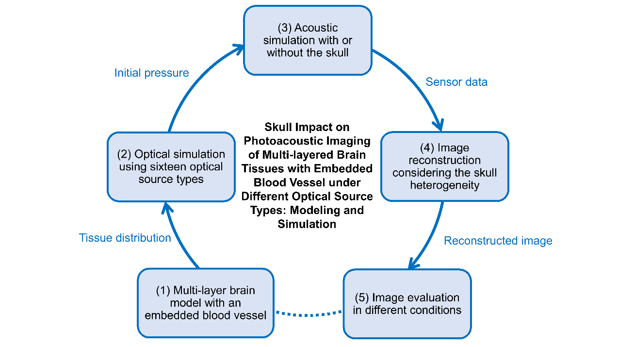
Figure 1: Schematic diagram of Modeling and simulation of multi-layer brain tissues embedded blood vessel by skull under different light source types.
Reference:
Please scan the QR code below to find more information:
Yang, X.; Chai, C.; Chen, Y.-H.; Sawan, M. Skull Impact on Photoacoustic Imaging of Multi-Layered Brain Tissues with Embedded Blood Vessel Under Different Optical Source Types: Modeling and Simulation. Bioengineering 2025, 12, 40. https://doi.org/10.3390/bioengineering12010040

Highlights:
1. Photoacoustic simulation and image reconstruction under 16 light sources have been established based on the entire process.
2. The influence of the skull on a multi-layered brain model with embedded blood vessels has been explored.
3. Quantitative analysis has been conducted on the characteristics of photoacoustic signals under different locations and model conditions.
Abstracts:
Skulls with high optical scattering and acoustic attenuation are a great challenge for photoacoustic imaging for human beings. To explore and improve photoacoustic generation and propagation, we conducted the photoacoustic simulation and image reconstruction of the multi-layer brain model with an embedded blood vessel under different optical source types. Based on the optical simulation results under different types of optical sources, we explored the characteristics of reconstructed images obtained from acoustic simulations with and without skull conditions.
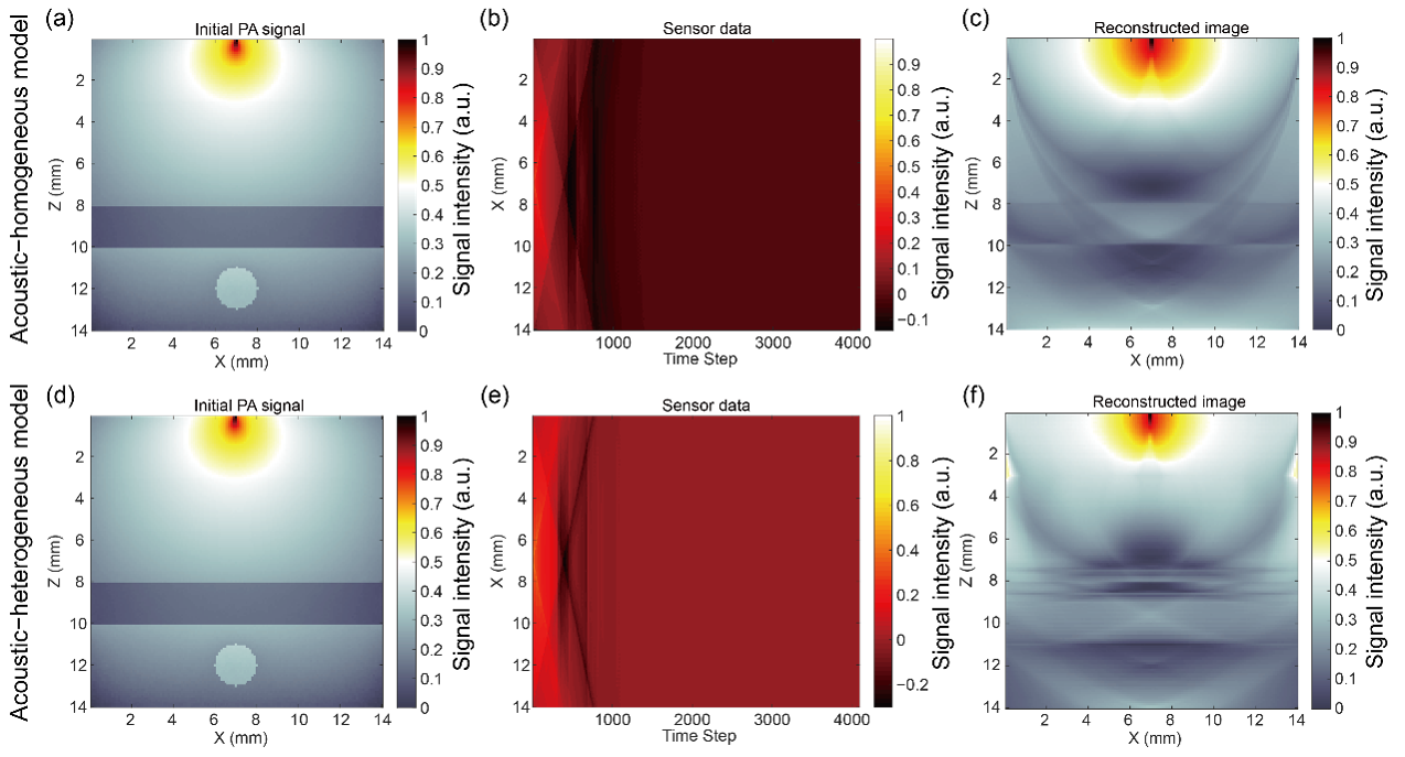
Figure 2: Simulation and reconstruction results of multi-layer brain tissues embedded with blood vessels under the illumination of a pencil beam.
Specifically, we focused on the detection of blood vessels and evaluated the image reconstruction features, morphological characteristics, and intensity of variations in the target vessels using optical and acoustic simulations. The results showed that under the initial photoacoustic signals, the types of optical source types corresponding to the strongest and weakest photoacoustic signals at different positions within the target region were consistent, while the optical source types were different in the reconstructed images. This study revealed the characteristics of acoustic signal transmission with and without skull conditions and its impact on image reconstruction. It further provides a theoretical basis for the selection of optical sources.
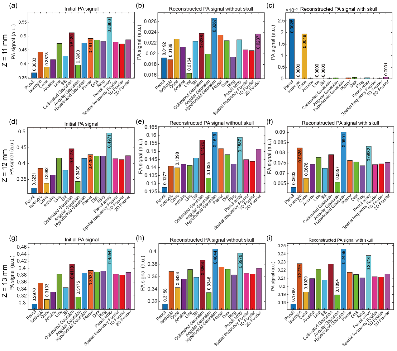
Figure 3: Maximum and minimum PA signals and corresponding optical source types at the specific position (x = 7 mm, z = 11, 12, and 13 mm) under the illumination of sixteen optical source types.