Titled “Monitoring Brain Activities Using fNIRS to Avoid Stroke”, this contribution has been published in IntechOpen, London. We present in this publication the fundamental of functional near-infrared spectroscopy (fNIRS) and the advantages of applying fNIRS for neuroimaging. In addition, we summarized the evolution of the fNIRS devices. In the last section, the applications of fNIRS to avoid stroke are presented. Congratulations to Yun-Hsuan Chen and to this book chapter’s co-author for the excellent achievement.
Citation
Y. Chen, and M. Sawan, "Monitoring Brain Activities Using fNIRS to Avoid Stroke", in Infrared Spectroscopy - Perspectives and Applications.London, United Kingdom: IntechOpen, 2022. Available: https://www.intechopen.com/online-first/82242
Abstract
Functional near-infrared spectroscopy (fNIRS) is an emerging wearable neuroimaging technique based on monitoring the hemodynamics of brain activity. First, the operation principle of fNIRS is described. This includes introducing the absorption spectra of the targeted molecule: the oxygenated and deoxygenated hemoglobin. Then, the optical path formed by emitters and detectors and the concentration of the molecules is determined using Beer-Lambert law. In the second part, the advantages of applying fNIRS are compared with other neuroimaging techniques, such as computed tomography and magnetic resonance imaging. The compared parameters include time and spatial resolution, immobility, etc. Next, the evolution of the fNIRS devices is shown. It includes the commercially available systems and the others under construction in academia. In the last section, the applications of fNIRS to avoid stroke are presented. The challenges of achieving good signal quality and high user comfort monitoring on stroke patients are discussed. Due to the wearable, user-friendly, and accessibility characteristics of fNIRS, it has the potential to be a complementary technique for real-time bedside monitoring of stroke patients. A stroke risk prediction system can be implemented to avoid stroke by combining the recorded fNIRS signals, routinely monitored physiological parameters, electronic health records, and machine learning models.
More information can be found at the following link:
https://doi.org/10.5772/intechopen.105461

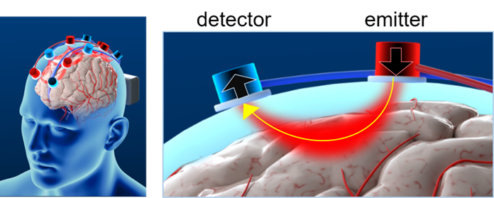
Fig.1: Placement of fNIRS optodes on the scalp. The hemoglobin changes in the blood vessels located in the optical path formed by a pair of emitter and detector can be monitored.


