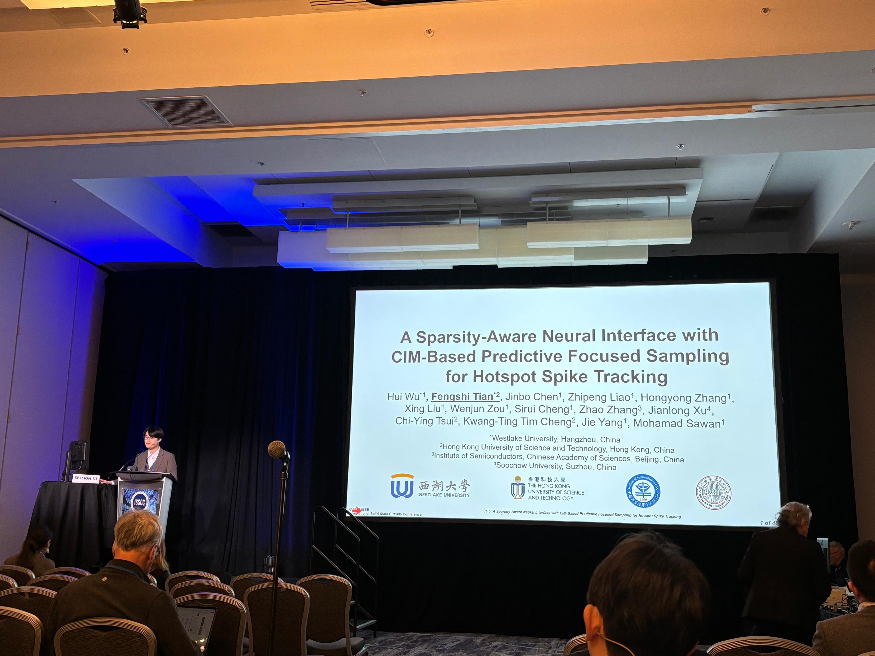
Big news! All six submitted papers from Professor Mohamad Sawan’s research center at Westlake University have been accepted by the 48th Annual International Conference of the IEEE Engineering in Medicine and Biology Society (IEEE EMBC).
This marks the third consecutive year that our team members have had papers accepted at this conference, and this year, we set a new record with the highest number of papers and a 100% acceptance rate.
Research Assistant Professor Dr. Fahimeh Marvi, postdoctoral fellow Dr. Hongyong Zhang, Ph.D students Xurong Gao, Chengpeng Chai, Kang Xiong, and research assistant Yuchen Sang will attend the conference in Toronto, Canada, this July.

(The following section provides a brief overview of the six accepted papers. Further details will be shared after the conference.)
#Paper 1
“Novel MEMS Platforms for Molecules-based Biosensing and Flexible Probing”
First Author:Fahimeh Marvi
Correspond Author:Mohamad Sawan
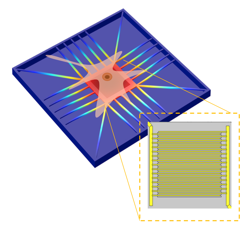
Figure 1. A schematic description of the proposed topology with representing the electrode part.
In this paper, we present a novel MEMS-based biosensing platform. Through optimized structures and a wet etching process, the fabricated devices achieve a mechanical sensitivity of 55 nm/kPa over a wide bandwidth of 50 kHz. The implemented BioMEMS devices are intended to enable the integration of optical and capacitive sensing techniques to simultaneously detect both biochemical and biophysical changes, making them suitable for applications such as noninvasive diagnostics and cell-based assays.
#Paper 2
“A Sustainable Exosomes-based Drug Delivery Technique Targeting Brain Cancer Therapy”
First Author:Hongyong Zhang
Correspond Author:Mohamad Sawan
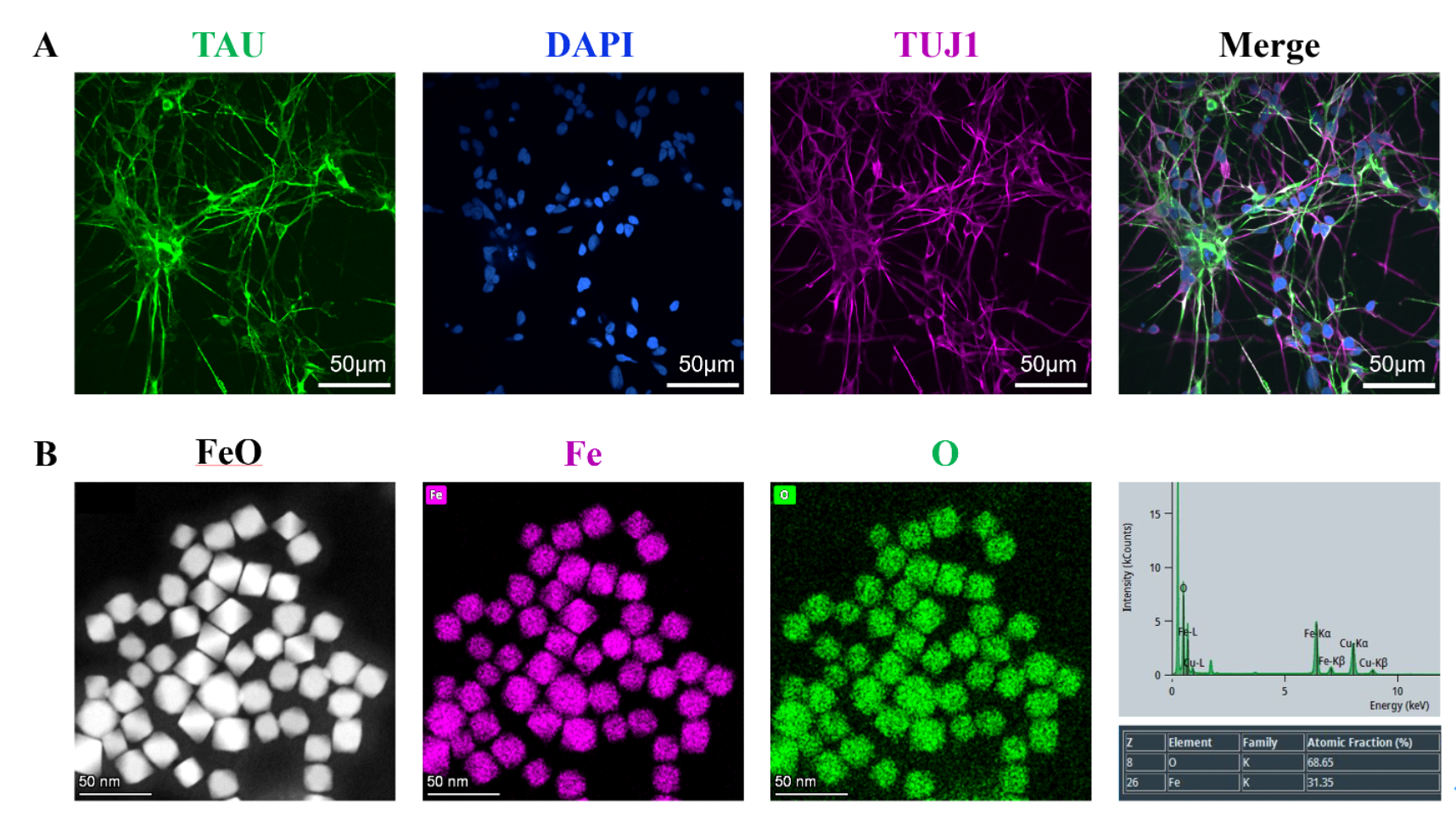
Figure 2. Characterization of neurons and MNPs: (A) Immunofluorescence staining of neurons; (B) TEM images of MNPs.
Exosome-based therapeutic strategies face challenges including complex purification procedures, limited production yield, and low drug loading efficiency. In this study, we proposed a method to sustainably produce exosomes by activating human neural cells with magnetic nanoparticles. The exosomes were then loaded with cisplatin and delivered to neural cancer cells, and magnetic manipulation was utilized to achieve targeted delivery, thereby reducing the side effects of chemotherapy.
#Paper 3
“Towards Effective Optogenetic-Based Vision Enhancement: Insights and Capabilities”
First Author:Xurong Gao
Correspond Author:Mohamad Sawan
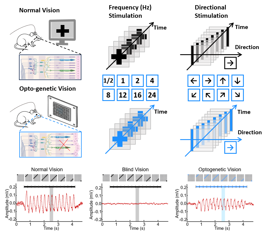
Figure 3. Schematic of the experimental setup for optogenetic vision mechanism research.
This study employs a mouse model, combining in vivo electrophysiological recordings with decoding techniques, to evaluate optogenetic vision at the cortical level. It advances optogenetic vision research from the ex vivo retina to the domain of cortical information processing and proposes a cortical decoding-based assessment framework for quantitatively evaluating and optimizing vision restoration strategies.
#Paper 4
“A MEG-Compatible Ultrasound Transducer with 1.5 nT Remanence”
First Author:Chengpeng Chai
Correspond Author:Mohamad Sawan
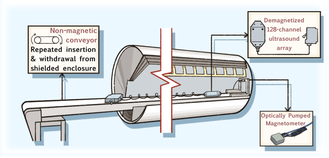
Figure 4. System-level concept of the OPM-compatible demagnetized ultrasound array and the magnetic cleanliness evaluation framework.
To acquire synchronized structural and neuromagnetic data for multimodal neuroimaging, strict requirements on magnetic cleanliness are imposed, which are difficult to meet with conventional medical imaging systems. To address this equipment–signal compatibility bottleneck, we present a high–magnetic-cleanliness ultrasound transducer based on conventional lead zirconate titanate (PZT) architecture and establish a reproducible residual-magnetism evaluation framework using OPMs, providing a critical engineering foundation for multimodal integration of ultrasound/photoacoustic imaging with OPM-MEG.
#Paper 5
“A Contactless Differential-capacitive Measurement of Brain Organoids”
First Author:Kang Xiong
Correspond Author:Mohamad Sawan
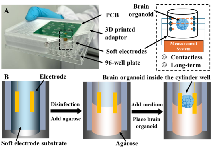
Figure 5. The schematic of brain organoid culture and monitor platform.
To avoid the issue that invasive operations may damage the development of brain organoids during monitoring, we propose a near-field wireless method based on capacitance measurement to assess the growth of brain organoids and design a custom differential capacitance measurement device for automated monitoring of organoid changes. The proposed method and device are expected to be applied in the biopharmaceutical industry to improve the efficiency of large-scale brain organoid culture monitoring.
#Paper 6
“Hemodynamic Activation In Sarcopenic Elderly During Hand Grip Tasks: An fNIRS Study”
Co-first Author:Liang Yi、Yuchen Sang
Co-correspond Author:Mohamad Sawan、Qin Zhang
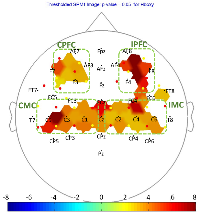
Figure 6. The SPM of HbO when comparing the activation between SG and CG. Darker red indicates that the concentration of HbO on SG is higher than that of CG.
Sarcopenia is the progressive loss of muscle mass and function. Commonly applied diagnostic techniques evaluate parameters measured directly from the muscle, providing limited information about cerebral neuronal activation. This study employed functional near-infrared spectroscopy (fNIRS) to monitor hand-grip tasks and found that sarcopenic patients required higher cerebral oxygenated hemoglobin activation to complete the tasks, along with significantly reduced functional connectivity between brain regions. fNIRS holds promise for distinguishing sarcopenia and monitoring neural activity.