Lower extremity motor dysfunction is a common consequence of stroke, traumatic brain injury, spinal cord injury, and other neurological disorders. Such impairments significantly compromise patients' independence in daily activities, reduce their quality of life, and place considerable burdens on families and healthcare systems. Moreover, motor deficits often coexist with fatigue, and their interaction further constrains rehabilitation endurance and recovery. However, how different movement types are modulated by the cortico‑muscular network in response to fatigue remains poorly understood.
A recent study addressing this issue has been published in IEEE Transactions on Neural Systems and Rehabilitation Engineering , a well‑known journal in the field of biomedical engineering and rehabilitation. Dr. Yuchen Xu, a former postdoctoral fellow at the CenBRAIN Neurotech Center of Excellence, Westlake University, is the first author, with Chair Professor Mohamad Sawan serving as the corresponding author.
This work systematically analyzed the changes in cortico‑cortical and cortico‑muscular network patterns during the concentric (T1: sit‑to‑stand) and eccentric (T2: stand‑to‑sit) phases of the sit‑to‑stand transition task, both before and after fatigue induction, with the aim of testing whether concentric and eccentric movements employ distinct fatigue‑compensation strategies. Elucidating these compensatory mechanisms may provide a neurophysiological basis for developing task‑dependent, phase‑specific rehabilitation approaches for patients with neurological injuries.
Abstract
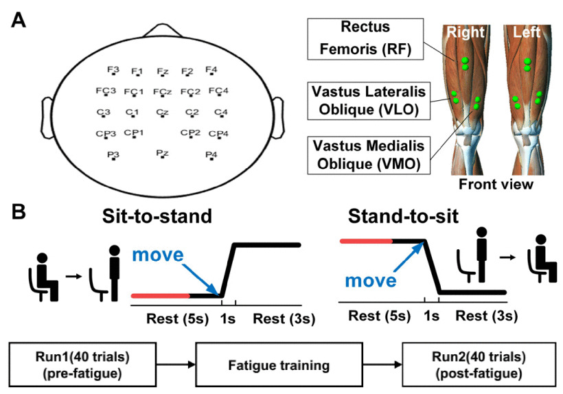
Fig. 1. (A) Placement of EEG and EMG recording electrodes. (B) Experimental paradigm consisting of pre-fatigue, fatigue and post-fatigue tasks. Each trial includes sit-to-stand and stand-to-sit tasks.
Ten healthy participants were recruited to perform sit‑to‑stand tasks—concentric (T1: sit‑to‑stand) and eccentric (T2: stand‑to‑sit)—under both pre‑fatigue and post‑fatigue conditions. During these tasks, electroencephalogram (EEG) and electromyogram (EMG) signals from the lower limbs were recorded. Muscle fatigue and mental fatigue were assessed via EMG mean power frequency (MPF) and EEG Lempel‑Ziv complexity (LZC), respectively. Inter‑cortical and cortico‑muscular networks were analyzed using generalized partial directed coherence (GPDC), with clustering coefficient (CC) and global efficiency (GE) as network metrics.
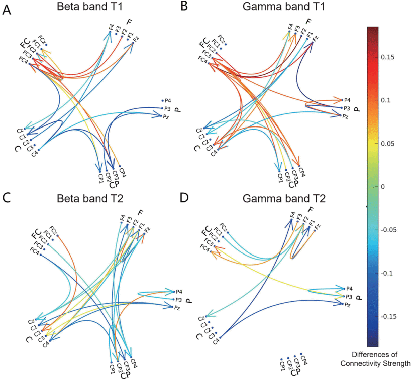
Fig. 2. The significant differences of connectivity strength between pre-fatigue and post-fatigue states (Post-fatigue–Pre-fatigue) in Beta band T1 (A), Gamma band T1 (B), Beta band T2 (C) and Gamma band T2 (D).
In the pre‑fatigue state, T1 exhibited stronger inter‑cortical connectivity than T2. In the post‑fatigue state, T1 further enhanced inter‑cortical connectivity, whereas T2 demonstrated significant increases in CC and GE within the beta band (P = 0.018 and P = 0.034, respectively). At the cortico‑muscular level, T1 shifted descending pathways from the beta to the gamma band, while T2 significantly upregulated both descending and ascending cortico‑muscular pathways in the beta and gamma bands.
These findings suggest that concentric movements rely more on compensatory inter‑cortical connectivity and band shifts within the cortico‑muscular network under fatigue, whereas eccentric movements exhibit greater inter‑cortical efficiency and stronger cortico‑muscular network strength. This study reveals distinct modulation patterns for concentric and eccentric movements in fatigued conditions, offering valuable insights for developing targeted rehabilitation training and therapeutic programs.
Reference
Y. Xu, C. Li, M. Wang, Y. -H. Chen, S. Zhang and M. Sawan, "Cortical and Cortico-Muscular Network Modulation Pattern of Eccentric and Concentric Movements in Fatigue State," in IEEE Transactions on Neural Systems and Rehabilitation Engineering, vol. 34, pp. 2762-2772, 2026.
More information can be found at the following link:
https://ieeexplore.ieee.org/document/11527007