Precise temperature monitoring is important for thermal ablation, photothermal therapy, focused ultrasound treatment, vascular physiology, and brain-function research. Photoacoustic imaging combines optical absorption contrast with ultrasonic detection depth, while its signal amplitude is inherently affected by temperature-dependent thermal and optical properties.
Conventional biomedical photoacoustic thermometry, however, commonly relies on hemoglobin-related wavelengths and may be influenced by hemoglobin concentration, oxygenation status, and local optical fluence. These factors are spatially and temporally heterogeneous in biological tissues and can be difficult to separate from the temperature response. Water is the most abundant component of soft tissue and has a relatively stable spatial distribution. Near 960 nm, its absorption exhibits a predictable temperature-dependent response associated with thermally induced changes in the hydrogen-bond network.
This study therefore investigated a water-sensitive photoacoustic thermometry strategy at 960 nm and evaluated it in human whole-blood cerebrovascular phantoms with and without a simplified bone-like, high-sound-speed shell.
To address the above issues, Professor Mohamad Sawan's center (CenBRAIN Neurotech) and Professor Liang Lei’s Lab at Westlake University proposed and validated a water-sensitive photoacoustic thermometry strategy based on 960 nm excitation.
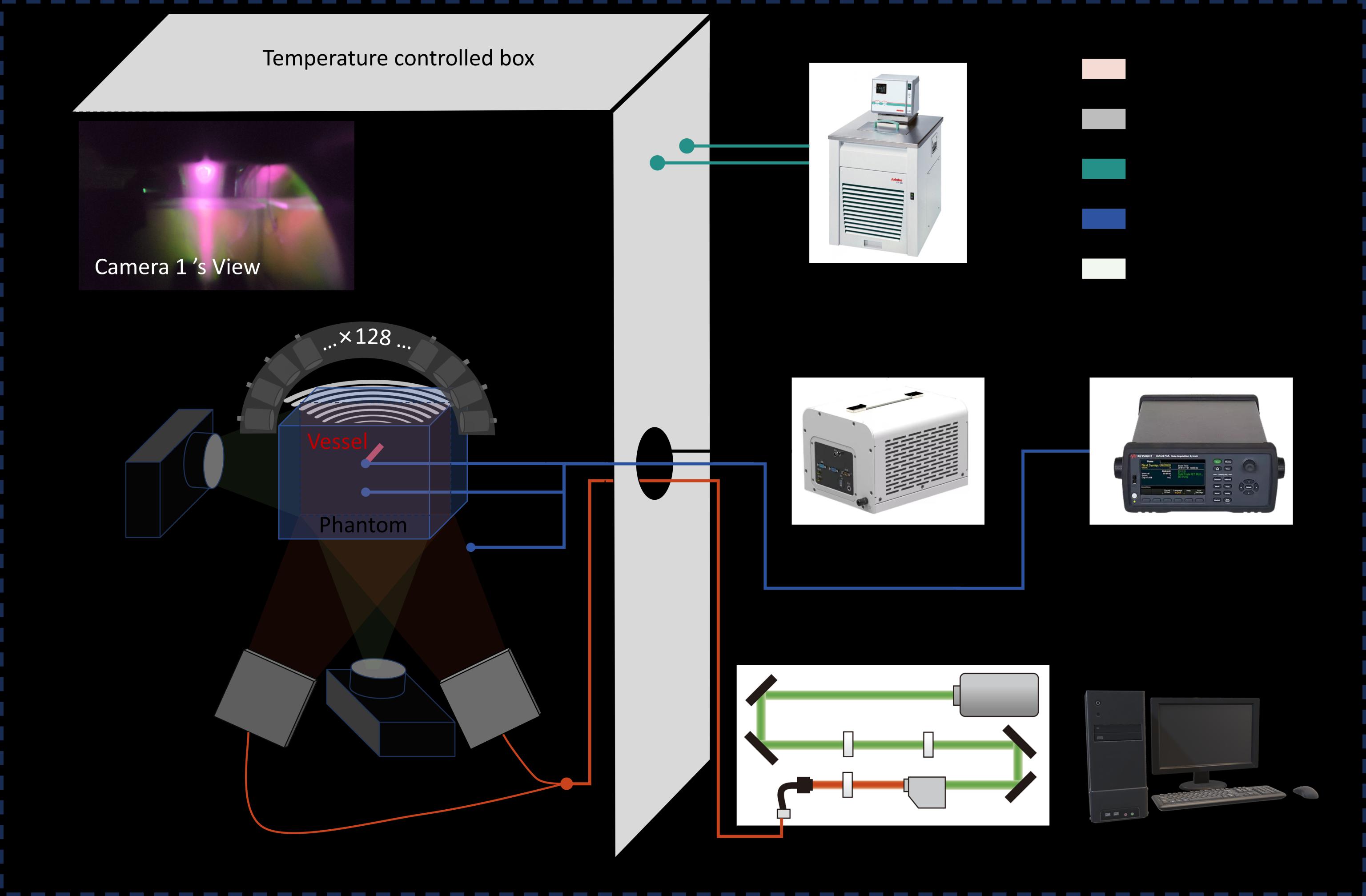
Fig. 1. Schematic of the photoacoustic temperature measurement system. The inset shows a camera-view photograph of the experimental setup inside the temperature-controlled box. The system consists of a temperature-controlled box, an excitation laser module, line-shaped optical illumination, a 128-element semi-ring ultrasound transducer array, a data acquisition unit, a temperature recording module, and a host computer for system control and data processing. The sample was illuminated by the pulsed excitation laser, and the generated photoacoustic signals were detected by the semi-ring ultrasound array and transferred to the acquisition system for image reconstruction and temperature-related analysis. Colored lines indicate the functional connections among the temperature-control, laser-excitation, acoustic-detection, signal-acquisition, and data-processing modules.
This work was published as a paper titled "Water-sensitive photoacoustic temperature characterization at 960 nm in cerebral vascular phantoms with CT co-registration" in the journal Photoacoustics.
Chengpeng Chai, a Ph.D student at CenBRAIN Neurotech, and Kaiyu Wang, a Ph.D student from Prof. Liang Lei’s lab, are co-first authors. The co-corresponding authors are Chair Professor Mohamad Sawanand Associate Professor Liang Lei. We thank the Young Elite Scientists Sponsorship Program for Doctoral Students of the China Association for Science and Technology, the Young Investigator Program of the Future Human Research Institute, the Research Fund for International Senior Scientists (RFISS), the Zhejiang Provincial Key R&D Program, the Zhejiang Provincial "Jianbing" and "Lingyan" R&D Program, and the Key Project of the Institute of Optoelectronics, Westlake University for their funding support.
Research Highlights
A water-absorption-dominated photoacoustic thermometry strategy, using 960 nm waveform, was proposed.
A phantom without a bone-like shell achieved an R2 of 0.996 and an RMSE of 0.08°C at a depth of 17.5 mm.
The proposed phantom remains maintaining an R2 > 0.976 and an RMSE of 0.13°C at a depth of 17.5 mm.
Paper Information
Chai C*, Wang K*, Chen L, Zhang J, Mao Y, Ren X, Li Z, Jin F, Chen YH, Lei L, Sawan M. Water-sensitive photoacoustic temperature characterization at 960 nm in cerebral vascular phantoms with CT co-registration. Photoacoustics. 2026 Jul 21; 51:100862.
https://www.sciencedirect.com/science/article/pii/S2213597926000686#ab0010
Abstract
This study proposed and validated a water-sensitive photoacoustic thermometry strategy based on 960 nm excitation. A laboratory-built photoacoustic computed tomography system equipped with a 128-element semi-ring ultrasound array was used to characterize agarose cerebrovascular phantoms containing a straight human whole-blood channel at a depth of approximately 17.5 mm. Measurements were performed from 35 to 40°C in phantom configurations with and without a 3D-printed SLA bone-like high-sound-speed shell.
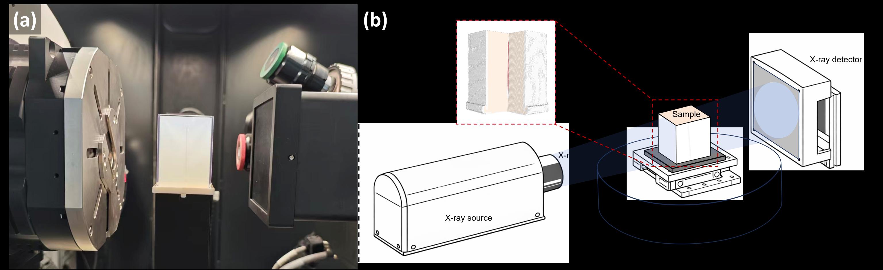
Fig. 2. CT imaging experiment of the phantom and its structural schematic. (a) Photograph of the phantom placed in the CT system; (b) schematic diagram of the CT imaging setup composed of the X-ray source, rotation stage, and detector.
Post-experimental CT imaging provided structural ground truth for rigid CT–PA registration, geometric scale correction, sound-speed adjustment, and structurally constrained ROI localization. The registration workflow achieved a mean target registration error of 0.3204 mm.
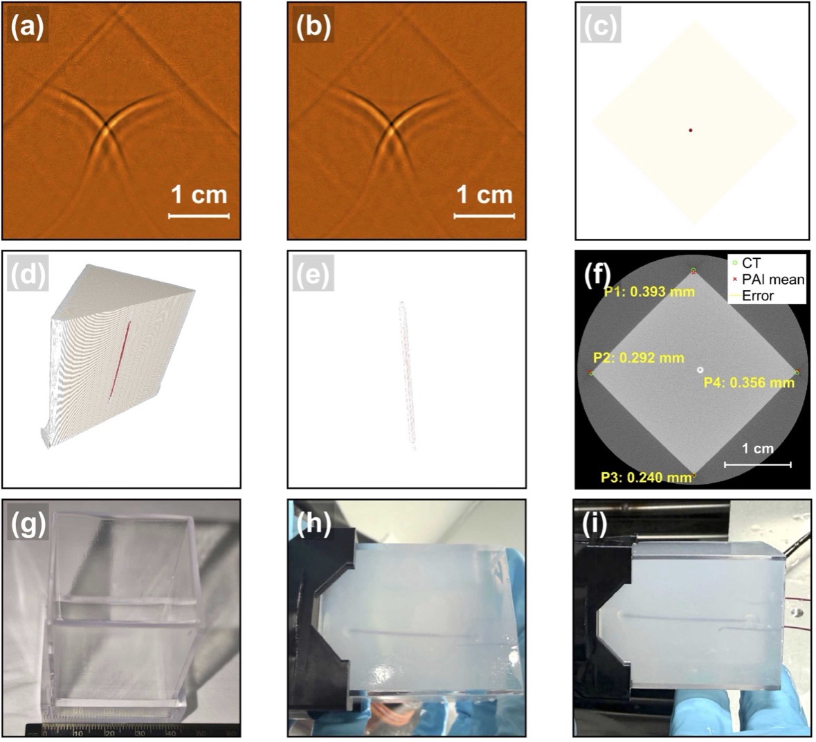
Fig. 3. CT-guided spatial validation and cross-modal registration in the bone-like shell phantom. (a) DAS photoacoustic reconstruction; (b) BP photoacoustic reconstruction. (c) CT-derived structure of the blood channel and PTFE tubing; (d) Longitudinal CT slice of the phantom; (e) Transverse CT slice of the phantom. (f) CT-PA landmark registration overlay showing CT landmarks, mean registered PA landmarks, and registration error vectors; (g) Photograph of the bone-like shell phantom with a scale bar; (h, i) Photographs of the phantom before and after the heating cycle, respectively, taken from the same view to show the macroscopic state of the phantom and the position of the thermocouple.
Three processing strategies were compared to evaluate frame-level behavior, averaging performance, and statistically aggregated inversion performance. Although the raw frame-wise signals retained an overall temperature-dependent trend, they exhibited considerable fluctuations, with an R² of 0.673 and a noise-equivalent temperature difference of 0.8079°C. After 100-frame averaging, R² increased to 0.989 and the noise-equivalent temperature difference decreased to 0.1240°C, demonstrating improved thermometric stability and sensitivity through temporal averaging.
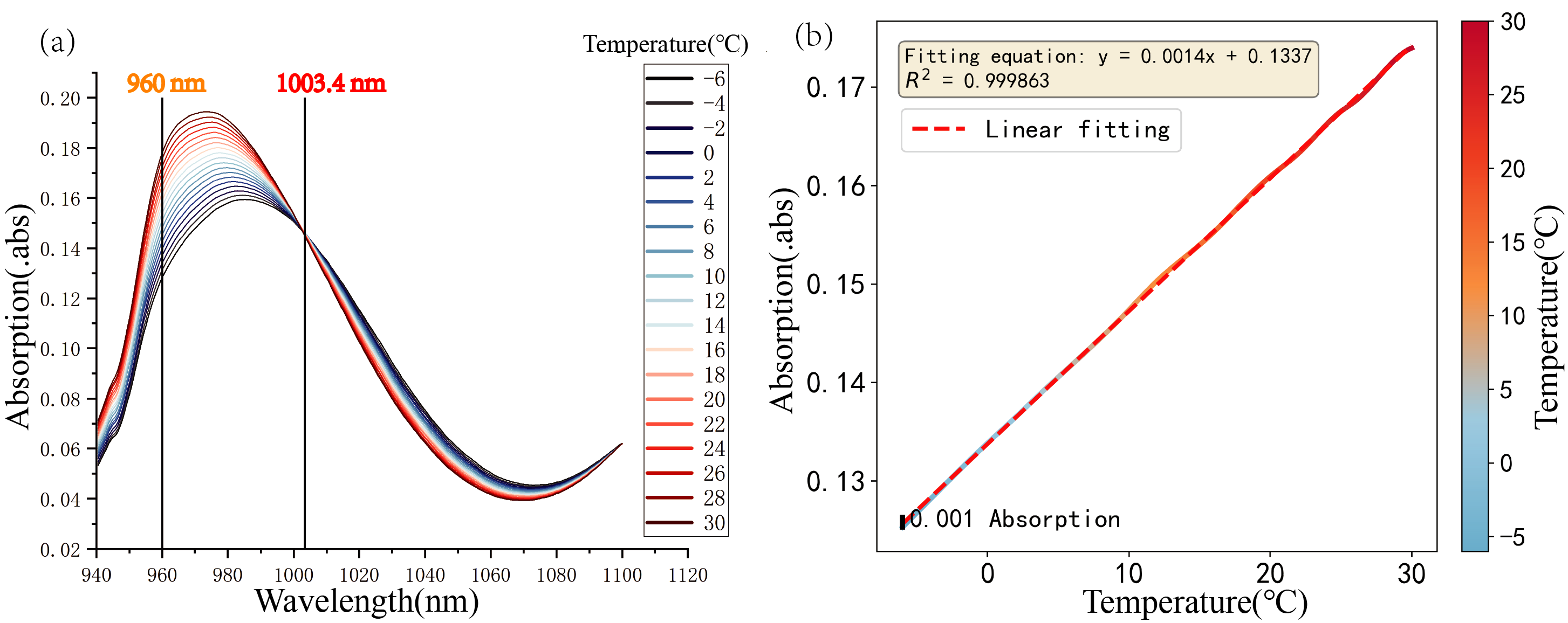
Fig. 4. Temperature-dependent absorption characteristics of ultrapure water. (a) Absorption spectra of ultrapure water in the 940-1120 nm band at different temperatures; (b) Relationship between absorbance and temperature at 960 nm.
Using 0.1°C temperature-bin median aggregation, R² further increased to 0.9957, with an apparent temperature inversion RMSE of 0.0773°C. In the phantom containing the bone-like shell, an R² above 0.976 and an apparent RMSE of 0.13°C were maintained. These RMSE values represent apparent inversion performance after statistical aggregation rather than single-frame absolute thermometric accuracy. Future incorporation of pulse-by-pulse optical-energy monitoring, normalization, and closed-loop laser-energy stabilization may reduce systematic fluctuations and improve single-frame and real-time thermometry.
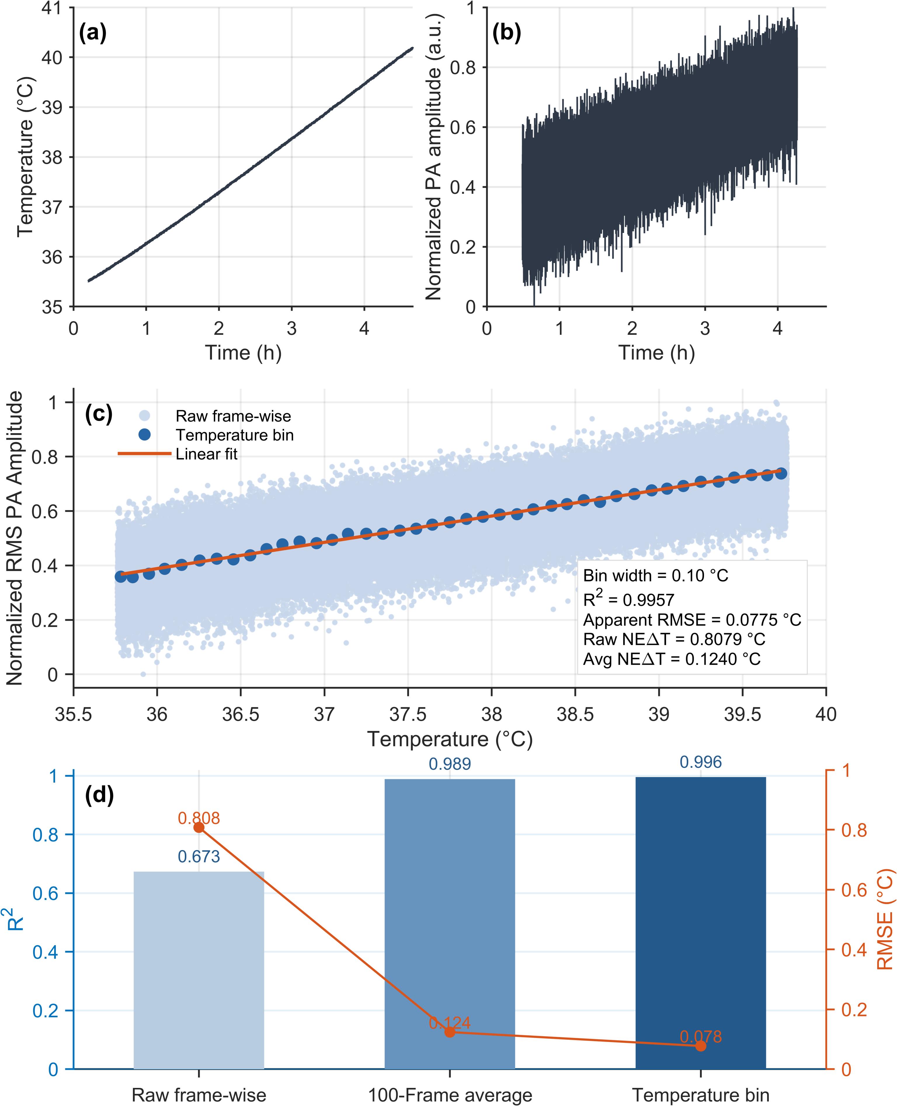
Fig. 5. Quantitative results of the temperature-related photoacoustic response in a cerebrovascular phantom without a bone-like shell. (a) Variation of the reference temperature over time during heating; (b) Variation of RMS photoacoustic amplitude within the selected ROI over time; (c) Relationship between temperature and ROI-based RMS photoacoustic amplitude, showing raw frame-wise data, 0.10°C temperature-binned median values, and the corresponding linear fit; the annotated NEΔT values represent the noise-equivalent temperature differences for raw frame-wise and 100-frame averaged analyses; (d) Comparison of R2 and apparent temperature inversion error (RMSE) under three statistical strategies: raw frame-by-frame, frame averaging, and temperature binning.